

Show condition progression and demonstrate the benefits of various treatment options
Now more than ever, we are hearing from Rendia customers that Exam Mode has become an invaluable tool in their practices. With its realistic, interactive, 3D views of the eye, it lets providers fly through the eye to explain anatomy, show condition progression and demonstrate the benefits of various treatment options. And Exam Mode can be seamlessly integrated into telehealth appointments, giving patients just as great an experience during a virtual visit as they would get in your office.
To see Exam Mode in action, play our Glaucoma One Minute Visit , which demonstrates how you can show a complex topic in just 60 seconds.
Below, we’re highlighting some of the treatments, views, and features that you might not know Exam Mode offers. Check out all the ways it can save provider time, enhance your patients’ experience and increase conversions.
Top 10 clips to show your patients
1. Thermal Pulsation – iLux – quickly show how the meibomian glands function, how they become blocked, and how you will target thermal pulsation to express the glands for dry eye relief.
2. MIGS treatments for Glaucoma – show the trabecular meshwork, how aqueous fluid cannot properly drain, and how during cataract surgery you can use micro-stents to effectively reduce intraocular pressure by creating a new pathway.
3. Laser Vitreolysis – Floaters – orient patients to the back of the eye, show how floaters develop, and how the procedure vaporizes the floaters to give patients relief from these bothersome bits.
4. Durysta (Alternative to Drops) – Glaucoma – show how a dissolvable ocular implant is inserted gently into the tissue to reduce eye pressure for several months.
5. DMEK – Fuchs’ Dystrophy – show corneal clouding, how vision becomes blurry, and how you’ll use microscopic incisions to replace just the endothelial layer of cells as compared to traditional corneal transplants which replaces the entire cornea, allowing for a faster visual recovery and fewer restrictions.
6. YAG – Cataract Surgery – ease concerns about a cataract “growing back” by showing the hazy capsule and how you will use a laser to make an opening in the center of the posterior capsule to let light pass through and improve vision.
7. Blephex – Demodex/Blepharitis – show the meibomian glands function, how they become blocked, and how BlephEx will clear out the collarettes and pathogens from the eyelid, similar to brushing your teeth.
8. Intacs – Keratoconus – show the normal shape of the cornea, how it changes as Keratoconus develops and how plastic ring inserts are placed in the outer areas of the cornea to change the curvature and reduce distortion.
9. Corneal Crosslinking – Keratoconus – show the normal shape of the cornea, how the shape changes as the links between corneal fibers weaken, and how you will strengthen the fibers and stabilize the shape of the cornea by administering drops of riboflavin and UV light.
10. DCR – Endoscopic – Tear Duct obstruction – demonstrate normal tear flow, how excessive tearing occurs when tears cannot properly drain due to a blockage, and how you will create a new drainage channel to bypass the blocked tear duct.
Exam Mode features
- Point-of-views (POVs) let you simulate how a patient’s vision will be affected as a condition progresses over time, motivating patients to be adherent, while helping family members understand the importance of monitoring and treatment. Thanks to the picture-in-picture layout, POVs let you simultaneously show the anatomy and visual simulation, and demonstrate how these change as the condition progresses over time. See how POVs work here.
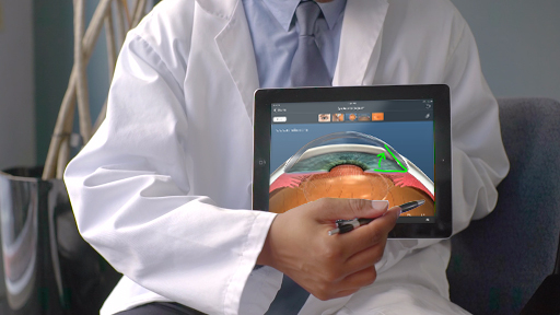
- The drawing tool allows you to draw on screen to highlight the most important anatomical structures, explain your surgical plan in greater detail, or simply impress your patient
- Exam Mode for iPad gives you the convenience of an app on a tablet to educate patients as you move between rooms, without the barrier of a large monitor between you. See how to use the touchscreen to navigate Exam Mode on an iPad.
- Hotspots are a fast way to navigate through the anatomy – you can click or tap to zoom in and out or rotate perspective, ensuring your patient understands exactly what you’re talking about. See how hotspots work.
What doctors say about Exam Mode
Doctors who use Exam Mode say, “It is a game changer in real-time patient education.” Joseph Deering, O.D., says he and his patients both love it. “Being able to quickly give them a tour of an eye to clear up some simple misconceptions leaps the conversation forward and personalizes their eye experience whether they are completely healthy, have advanced disease, are 5 or are 100.”
Lance Kugler, M.D., medical director of Kugler Vision in Omaha, Nebraska, found that Exam Mode is a great solution for virtual patient education. “In a telemedicine situation, a 2D physical eye model is not very valuable. It’s more effective to use a tool like Rendia’s Exam Mode that’s optimized for a screen.”
Learn how Dr. Paul Karpecki uses Exam Mode to explain glaucoma and MIGS to patients with glaucoma and cataracts. Watch his 4-minute tutorial here.